
Tập tin:GFP Superresolution Christoph Cremer.JPG
GFP_Superresolution_Christoph_Cremer.JPG (538×389 điểm ảnh, kích thước tập tin: 156 kB, kiểu MIME: image/jpeg)
Tập tin này từ Wikimedia Commons. Trang miêu tả nó ở đấy được sao chép dưới đây. Commons là kho lưu trữ tập tin phương tiện có giấy phép tự do. Bạn có thể tham gia. |
Miêu tả
| Miêu tả |
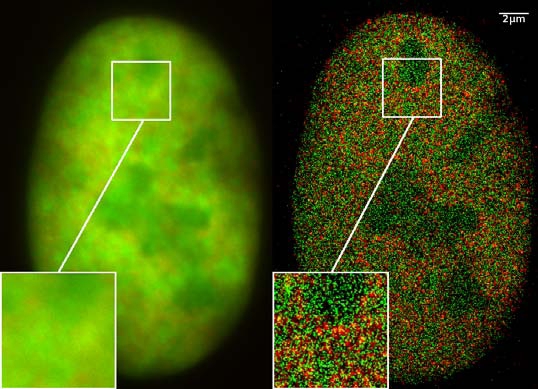
GFP superresolution, optical nanoscopy ( Christoph Cremer, emeritus at Heidelberg university [1]) View of a nucleus of a bone cancer cell: using normal high resolution fluorescence microscopy, it is not possible to distinguish details of its structure (image on the left). Using the two Color Localization Microscopy 2CLM (image on the right) it is possible to localize 70,000 histone molecules (red: RFP-H2A) and 50,000 chromatin remodeling proteins (green: GPF-Snf2H) in a field of view of 470 µm2 with an optical depth of 600 nm. Common fluorescence markers were used. 2CLM is the only optical nanoscopy method that allows position based co-localization of single molecules at high density in a wide field of view using conventional fluorescent proteins such as GFP, YFP, RFP, or other conventional fluorochromes. Due to its high optical single molecule resolution, 2CLM allows significantly more precise analyses of potential protein interactions than FRET-(Fluorescence Resonance Energy Transfer) technology, which is at present the preferred method for such investigations. This is of particular significance in studies of biomolecular machines (BMMs) within cells: Single BMMS can be analysed, including the number of molecules of a given type; distances between proteins in these BMMs often are substantially greater than those that can be analyzed by FRET (restricted to a maximum distance of only a few nm). Possible to use conventional, well established and inexpensive fluorescent dyes, from the GFP group, and its dye variants, to the well-known Alexa and fluorescein dyes. Fundamental to SPDMphymod are blinking phenomena (flashes of fluorescence), induced by reversible bleaches (metastable dark states). Individual molecules of the same spectral emission color can be detected. Publikation: Manuel Gunkel, Fabian Erdel, Karsten Rippe, Paul Lemmer, Rainer Kaufmann, Christoph Hörmann, Roman Amberger and Christoph Cremer: Dual color localization microscopy of cellular nanostructures. In: Biotechnology Journal, 2009, 4, 927-938. ISSN 1860-6768 |
| Ngày | 073009 |
| Nguồn gốc | Tác phẩm được tạo bởi người tải lên |
| Tác giả | Andy Nestl |
| Giấy phép (Dùng lại tập tin) |
Gallery
- Super Resolution Microscopy - Localisation Microscopy
-
 Breast Cancer Cells: 3D Dual Color Super Resolution Microscopy of Her2 and Her3 & cluster calculations
 -
 Single YFP molecule detection in a human cancer cell. Typical distance measurements 15 nm
 -
 Co- localisation microscopy with GFP and RFP fusion proteins (nucleus of a bone cancer cell) 120.000 localized molecules in a widefield area(470 µm2)
 -
 Label-free Localisation Microscopy SPDM - Super Resolution Microscopy reveals prior undetebable intracellular structures
 -
 Investigation of human eye tissue, affected by macular degeneration AMD
 -
 Virus Super Resolution Microscopy SPDM Cremer/Wege labs


Giấy phép
- Bạn được phép:
- chia sẻ – sao chép, phân phối và chuyển giao tác phẩm
- pha trộn – để chuyển thể tác phẩm
- Theo các điều kiện sau:
- ghi công – Bạn phải ghi lại tác giả và nguồn, liên kết đến giấy phép, và các thay đổi đã được thực hiện, nếu có. Bạn có thể làm các điều trên bằng bất kỳ cách hợp lý nào, miễn sao không ám chỉ rằng người cho giấy phép ủng hộ bạn hay việc sử dụng của bạn.
- chia sẻ tương tự – Nếu bạn biến tấu, biến đổi, hoặc làm tác phẩm khác dựa trên tác phẩm này, bạn chỉ được phép phân phối tác phẩm mới theo giấy phép y hệt hoặc tương thích với tác phẩm gốc.

|
Bạn có quyền sao chép, phân phối và/hoặc sửa đổi tài liệu này theo những điều khoản được quy định trong Giấy phép Tài liệu Tự do GNU, phiên bản 1.2 hoặc các phiên bản mới hơn được Quỹ Phần mềm Tự do; quy định; ngoại trừ những phần không được sửa đổi, bìa trước và bìa sau. Bạn có thể xem giấy phép nói trên ở phần Giấy phép Tài liệu Tự do GNU. |
Miêu tả
- ↑ https://www.physik.uni-heidelberg.de/personen/lsf.php?details=1537 |titel=Fakultät für Physik und Astronomie |abruf=2020-10-01
Lịch sử tập tin
Nhấn vào ngày/giờ để xem nội dung tập tin tại thời điểm đó.
| Ngày/giờ | Hình xem trước | Kích cỡ | Thành viên | Miêu tả | |
|---|---|---|---|---|---|
| hiện tại | 12:14, ngày 30 tháng 7 năm 2009 | | 538×389 (156 kB) | Andy Nestl | {{Information |Description=GFP superresolution, optical nanoscopy (Christoph Cremer) |Source=Own work by uploader |Date=073009 |Author=Andy Nestl |Permission=given by Christoph Cremer, University of Heidelberg |other_versions= }} |
Trang sử dụng tập tin
Sử dụng tập tin toàn cục
Những wiki sau đang sử dụng tập tin này:
- Trang sử dụng tại ar.wikipedia.org
- Trang sử dụng tại be.wikipedia.org
- Trang sử dụng tại bn.wikipedia.org
- Trang sử dụng tại ca.wikipedia.org
- Trang sử dụng tại cs.wikipedia.org
- Trang sử dụng tại de.wikipedia.org
- Trang sử dụng tại en.wikipedia.org
- Trang sử dụng tại en.wikibooks.org
- Trang sử dụng tại eo.wikipedia.org
- Trang sử dụng tại fa.wikipedia.org
- Trang sử dụng tại fr.wikipedia.org
- Trang sử dụng tại gl.wikipedia.org
- Trang sử dụng tại he.wikipedia.org
- Trang sử dụng tại it.wikipedia.org
- Trang sử dụng tại mai.wikipedia.org
- Trang sử dụng tại ne.wikipedia.org
- Trang sử dụng tại nl.wikipedia.org
- Trang sử dụng tại pl.wikipedia.org
- Trang sử dụng tại pt.wikipedia.org
- Trang sử dụng tại sv.wikipedia.org
- Trang sử dụng tại ta.wikipedia.org
- Trang sử dụng tại zh.wikipedia.org