
Tập tin:Anatomy of Human Ear with Cochlear Frequency Mapping.svg

Kích thước bản xem trước PNG này của tập tin SVG: 674×519 điểm ảnh. Độ phân giải khác: 312×240 điểm ảnh | 624×480 điểm ảnh | 998×768 điểm ảnh | 1.280×986 điểm ảnh | 2.560×1.971 điểm ảnh.
Tập tin gốc (tập tin SVG, 674×519 điểm ảnh trên danh nghĩa, kích thước: 33 kB)
Tập tin này từ Wikimedia Commons. Trang miêu tả nó ở đấy được sao chép dưới đây. Commons là kho lưu trữ tập tin phương tiện có giấy phép tự do. Bạn có thể tham gia. |
Miêu tả
| Miêu tả |
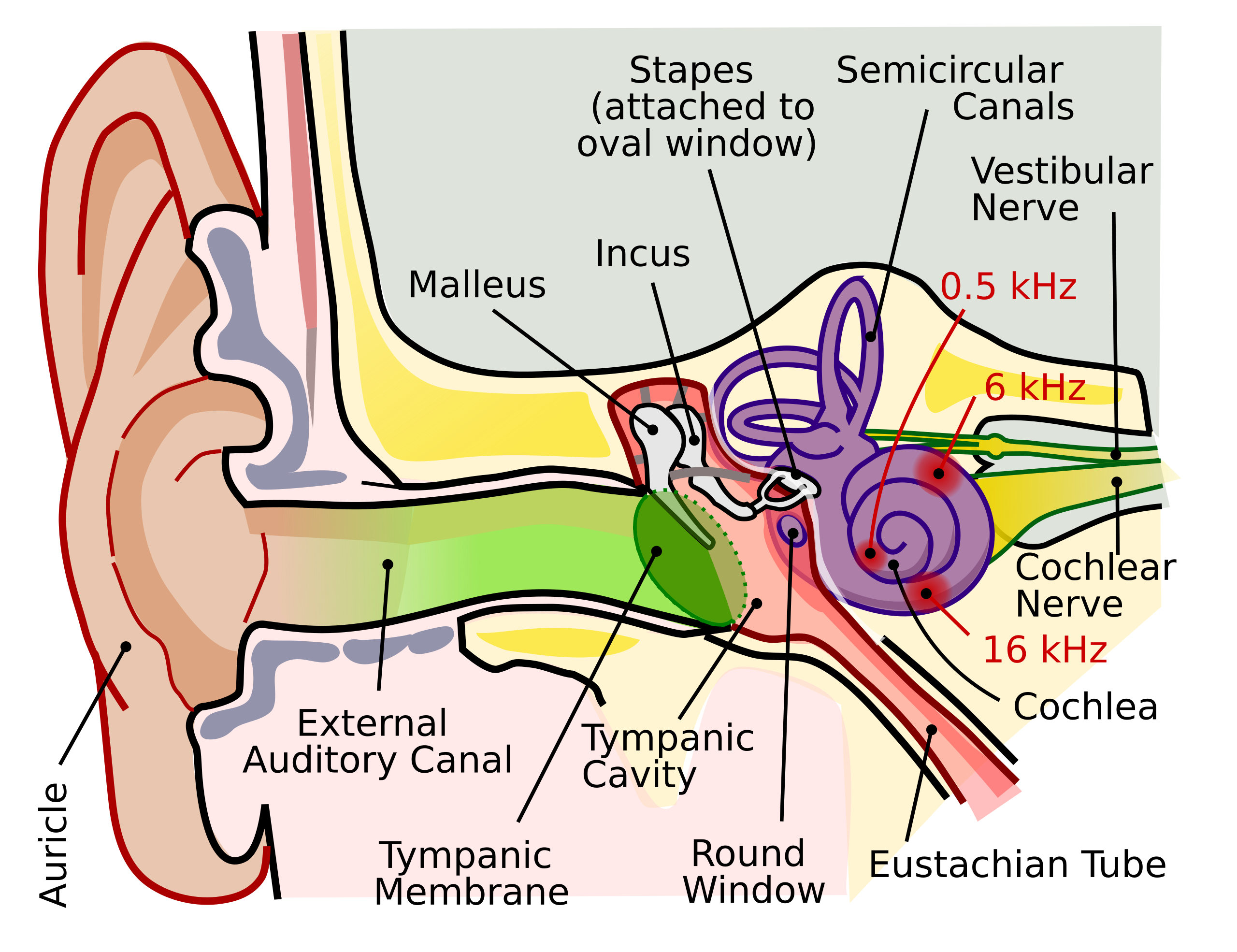
English: The human ear and frequency mapping in the cochlea. The three ossicles incus, malleus, and stapes transmit airborne vibration from the tympanic membrane to the oval window at the base of the cochlea. Because of the mechanical properties of the basilar membrane within the snail-shaped cochlea, high frequencies will produce a vibration peak near the oval window, whereas low frequencies will stimulate receptors near the apex of the cochlea (locations for three frequencies indicated schematically). Information from the cochlear receptor cells is transmitted to the cochlear nuclei via the 8th cranial nerve, and on through the midbrain to the cortex. |
| Ngày | |
| Nguồn gốc | Tác phẩm được tạo bởi người tải lên (Original text: Own work by uploader, derived from File:Anatomy_of_the_Human_Ear.svg) |
| Tác giả | Inductiveload |
| Giấy phép (Dùng lại tập tin) |
Tập tin này được phát hành theo Giấy phép Creative Commons Ghi công–Chia sẻ tương tự 2.5 Chung
|
| Phiên bản khác |
[]
|
| SVG genesis | This vector image was created with Inkscape. This file is translated using SVG switch elements: all translations are stored in the same file. |


Lịch sử tập tin
Nhấn vào ngày/giờ để xem nội dung tập tin tại thời điểm đó.
| Ngày/giờ | Hình xem trước | Kích cỡ | Thành viên | Miêu tả | |
|---|---|---|---|---|---|
| hiện tại | 21:29, ngày 16 tháng 9 năm 2018 | | 674×519 (33 kB) | JoKalliauer | added systemLanguage="eo" |
| 17:21, ngày 16 tháng 9 năm 2018 |  | 674×519 (32 kB) | JoKalliauer | added systemLanguage="de" | |
| 05:33, ngày 11 tháng 9 năm 2018 |  | 674×519 (87 kB) | Jmarchn | Bigger (proportional real size) and full redraw (more realistic) of the auricle. Ossicles in white colour. Eardrum with contour. Added 3 labels. Add fundus to the bone and subcutaneous tissues, add superior auricular muscle, add transparency to middle ear, add separation between middle and inner ear, add division to internal auditory canal. | |
| 13:40, ngày 29 tháng 4 năm 2009 |  | 800×600 (98 kB) | Inductiveload | swap incus/malleus | |
| 15:10, ngày 15 tháng 2 năm 2009 |  | 800×600 (98 kB) | Inductiveload | {{Information |Description={{en|1=The human ear and frequency mapping in the cochlea. The three ossicles incus, malleus, and stapes transmit airborne vibration from the tympanic membrane to the oval window at the base of the cochlea. Because of the mechan |
Trang sử dụng tập tin
Chưa có trang nào ở Wikipedia tiếng Việt liên kết đến tập tin này.
Sử dụng tập tin toàn cục
Những wiki sau đang sử dụng tập tin này:
- Trang sử dụng tại en.wikipedia.org
- Trang sử dụng tại en.wikibooks.org
- Trang sử dụng tại eo.wikipedia.org
- Trang sử dụng tại he.wikipedia.org
- Trang sử dụng tại lt.wikipedia.org
- Trang sử dụng tại www.wikidata.org