
Tập tin:Nematocyst discharge.png
Không có độ phân giải cao hơn.
Nematocyst_discharge.png (480×371 điểm ảnh, kích thước tập tin: 190 kB, kiểu MIME: image/png)
Tập tin này từ Wikimedia Commons. Trang miêu tả nó ở đấy được sao chép dưới đây. Commons là kho lưu trữ tập tin phương tiện có giấy phép tự do. Bạn có thể tham gia. |

|
Hình ảnh thuộc thể loại "biology" cần được vẽ lại bằng đồ họa vector theo định dạng tập tin SVG. Để biết ưu điểm của định dạng này, hãy đọc Commons:Media for cleanup. Nếu tập tin này đã có SVG, xin hãy tải lên đây rồi thay bản mẫu này bằng {{vector version available|tên hình mới.svg}}.
|
Miêu tả
| Miêu tả |
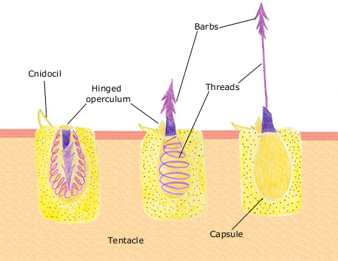
English: The diagram above shows the anatomy of a nematocyst cell and its “firing” sequence, from left to right. On the far left is a nematocyst inside its cellular capsule. The cell’s thread is coiled under pressure and wrapped around a stinging barb. When potential prey makes contact with the tentacles of a polyp, the nematocyst cell is stimulated. This causes a flap of tissue covering the nematocyst—the operculum—to fly open. The middle image shows the open operculum, the rapidly uncoiling thread and the emerging barb. On the far right is the fully extended cell. The barbs at the end of the nematocyst are designed to stick into the polyp’s victim and inject a poisonous liquid. When subdued, the polyp’s tentacles move the prey toward its mouth and the nematocysts recoil back into their capsules. |
| Ngày | 11 tháng 4 năm 2007 (ngày tải lên ban đầu) |
| Nguồn gốc | Chuyển từ en.wikipedia sang Commons. |
| Tác giả | The original uploader was Spaully tại Wikipedia Tiếng Anh. |
Giấy phép
This file is licensed under Creative Commons ShareAlike 1.0 License.
Creative Commons has retired this legal tool and does not recommend that it be applied to works.
|
Hình này thuộc phạm vi công cộng tại Hoa Kỳ vì nó là của Cục Quản lý Khí quyển và Đại dương Quốc gia Hoa Kỳ, được tạo ra trong quá trình công tác chính thức của nhân viên.
|
Nhật trình tải lên đầu tiên
Trang miêu tả gốc từng tồn tại ở đây. Tất cả các tên người dùng sau là tên người dùng tại en.wikipedia.
- 2007-04-11 17:10 Spaully 480×371×8 (194868 bytes) Modified from: http://www.oceanservice.noaa.gov/education/kits/corals/media/supp_coral01b.html {{Information |Description=Nematocyst discharge process. |Source= Modified from [http://www.oceanservice.noaa.gov/education/kits/corals/media/supp_coral01b.html
Lịch sử tập tin
Nhấn vào ngày/giờ để xem nội dung tập tin tại thời điểm đó.
| Ngày/giờ | Hình xem trước | Kích cỡ | Thành viên | Miêu tả | |
|---|---|---|---|---|---|
| hiện tại | 17:29, ngày 13 tháng 10 năm 2007 | | 480×371 (190 kB) | Alison | {{Information |Description===Description== The diagram above shows the anatomy of a nematocyst cell and its “firing” sequence, from left to right. On the far left is a nematocyst inside its cellular capsule. The cell’s thread is coiled under pressur |
Trang sử dụng tập tin
Có 1 trang tại Wikipedia tiếng Việt có liên kết đến tập tin (không hiển thị trang ở các dự án khác):
Sử dụng tập tin toàn cục
Những wiki sau đang sử dụng tập tin này:
- Trang sử dụng tại ca.wikipedia.org
- Trang sử dụng tại ceb.wikipedia.org
- Trang sử dụng tại en.wikipedia.org
- Trang sử dụng tại fr.wikipedia.org
- Trang sử dụng tại hr.wikipedia.org
- Trang sử dụng tại id.wikipedia.org
- Trang sử dụng tại it.wikibooks.org
- Trang sử dụng tại ja.wikipedia.org
- Trang sử dụng tại lv.wikipedia.org
- Trang sử dụng tại ms.wikipedia.org
- Trang sử dụng tại my.wikipedia.org
- Trang sử dụng tại pa.wikipedia.org
- Trang sử dụng tại pt.wikipedia.org
- Trang sử dụng tại simple.wikipedia.org
- Trang sử dụng tại sv.wikipedia.org
- Trang sử dụng tại te.wikipedia.org
- Trang sử dụng tại th.wikipedia.org