Thành viên:Naazulene/Cơ ức đòn chũm
| Sternocleidomastoid | |
|---|---|
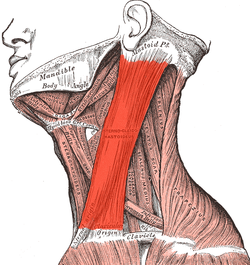
 Neck muscles, with the sternocleidomastoid in dark red | |
 The sternocleidomastoid (right muscle shown) can be clearly observed when rotating the head. | |
| Chi tiết | |
| Phát âm | (/ˌstɜːrnoʊˌklaɪdəˈmæsˌtɔɪd, |
| Nguyên ủy | Manubrium and medial portion of the clavicle |
| Bám tận | Mastoid process of the temporal bone, superior nuchal line |
| Động mạch | Occipital artery and the superior thyroid artery |
| Dây thần kinh | Motor: spinal accessory nerve sensory: cervical plexus Proprioceptive: ventral rami of C2-3 |
| Hoạt động | Unilaterally: contralateral cervical rotation, ipsilateral cervical flexion Bilaterally: cervical flexion, elevation of sternum and assists in forced inhalation. |
| Định danh | |
| Latinh | musculus sternocleidomastoideus |
| Thuật ngữ giải phẫu của cơ | |
Cơ ức đòn chũm (hay cơ ức - đòn - chũm, tiếng Anh: sternocleidomastoid muscle, tiếng Pháp: Le muscle sterno-cléido-mastoïdien) s là một trong nhữn m cơ lớn và nông (gần da) nhất của cơ thể người. Cơ này năm hai bên cổ. Hoạt động cơ bản của cơ này là xoay đầu về phía đối diện (khi một cơ hoạt động), và gập cổ - hay gật đầu (khi hai cơ cùng hoạt động). Cơ này nối với dây thần kinh phụ.
Từ nguyên và vị trí[sửa | sửa mã nguồn]
Cơ ức đòn chũm bắt đầu từ chuôi xương ức và xương đòn, bám tận đến mõm chũm của xương thái dương thuộc sọ.
Cấu trúc[sửa | sửa mã nguồn]
Cơ ức đòn chũm có nguyên ủy ở hai vị trí: chuôi xương ức và xương đòn.[3] Nó kéo dài theo hướng chéo qua cổ và bám tận vào mõm chũm của xương thái dương thuộc sọ, bằng một lớp cân mỏng.[3][4] Cơ ức đòn chũm dày và hẹp ở đoạn giữa mỏng và rộng dần về hai đầu.
Đầu phía xương đòn là một bó cơ tròn, nhiều gân về trước, nhiều thịt về sau, có nguyên ủy là phần trên của bề trước của chuôi xương ức (manubrium sterni). Nó đi dọc lên trên và chéo ra sau.
Đầu phía xương đòn được cấu tạo từ những sợi cân và thịt, có nguyên ủy là bề trên, trước của phần ba giữa của xương đòn; hướng của nó gần như thẳng đứng.
Hai đầu này được tách biệt ở nguyên ủy bởi một khoảng hình tam giác (gọi là lesser supraclavicular fossa), nhưng chúng dần hòa vào nhau dưới khoảng giữa của cổ, thành một bó cơ duy nhất dày và tròn, bám tận bằng một màng gân mạnh vào bề bên của mõm chũm (từ đỉnh đến rìa trên), và bởi một màng cân mỏng vào bán phần bên của đường gáy trên của xương chẩm.

Nerve supply[sửa | sửa mã nguồn]
Cơ ức đòn chũm, được gắn vào dây thần kinh phụ của mỗi bên. Nó chỉ cung cấp sợi thần kinh vận động.
The sternocleidomastoid is innervated by accessory nerve of the same side.[5][6] It supplies only motor fibres. The cervical plexus supplies sensation, including proprioception, from the ventral primary rami of C2 and C3.[5]
Variation[sửa | sửa mã nguồn]
The clavicular origin of the sternocleidomastoid varies greatly: in some cases the clavicular head may be as narrow as the sternal; in others it may be as much as 7,5 milimét (0,30 in)[chuyển đổi: số không hợp lệ] in breadth.
When the clavicular origin is broad, it is occasionally subdivided into several slips, separated by narrow intervals. More rarely, the adjoining margins of the sternocleidomastoid and trapezius are in contact. This would leave no posterior triangle.
The supraclavicularis muscle arises from the manubrium behind the sternocleidomastoid and passes behind the sternocleidomastoid to the upper surface of the clavicle.
Function[sửa | sửa mã nguồn]
The function of this muscle is to rotate the head to the opposite side or obliquely rotate the head.[3] It also flexes the neck.[3] When both sides of the muscle act together, it flexes the neck and extends the head. When one side acts alone, it causes the head to rotate to the opposite side and flexes laterally to the same side (ipsilaterally).
It also acts as an accessory muscle of respiration, along with the scalene muscles of the neck.
Contraction[sửa | sửa mã nguồn]
The signaling process to contract or relax the sternocleidomastoid begins in Cranial Nerve XI, the accessory nerve. The accessory nerve nucleus is in the anterior horn of the spinal cord around C1-C3, where lower motor neuron fibers mark its origin. The fibers from the accessory nerve nucleus travel upward to enter the cranium via the foramen magnum. The internal carotid artery to reach both the sternocleidomastoid muscles and the trapezius. After a signal reaches the accessory nerve nucleus in the anterior horn of the spinal cord, the signal is conveyed to motor endplates on the muscle fibers located at the clavicle. Acetylcholine (ACH) is released from vesicles and is sent over the synaptic cleft to receptors on the postsynaptic bulb. The ACH causes the resting potential to increase above -55mV, thus initiating an action potential which travels along the muscle fiber. Along the muscle fibers are t-tubule openings which facilitate the spread of the action potential into the muscle fibers. The t-tubule meets with the sarcoplasmic reticulum at locations throughout the muscle fiber, at these locations the sarcoplasmic reticulum releases calcium ions that results in the movement of troponin and tropomyosin on thin filaments. The movement of troponin and tropomyosin is key in facilitating the myosin head to move along the thin filament, resulting in a contraction of the sternocleidomastoid muscle.[7]
Anatomical landmark[sửa | sửa mã nguồn]

The sternocleidomastoid is within the investing fascia of the neck, along with the trapezius muscle, with which it shares its nerve supply (the accessory nerve). It is thick and thus serves as a primary landmark of the neck, as it divides the neck into anterior and posterior cervical triangles (in front and behind the muscle, respectively) which helps define the location of structures, such as the lymph nodes for the head and neck.[8]
Many important structures relate to the sternocleidomastoid, including the common carotid artery, accessory nerve, and brachial plexus.
Clinical significance[sửa | sửa mã nguồn]
Examination of the sternocleidomastoid muscle forms part of the examination of the cranial nerves. It can be felt on each side of the neck when a person moves their head to the opposite side.[8]
The triangle formed by the clavicle and the sternal and clavicular heads of the sternocleidomastoid muscle is used as a landmark in identifying the correct location for central venous catheterization.[cần nguồn y khoa]
Contraction of the muscle gives rise to a condition called torticollis or wry neck, and this can have a number of causes. Torticollis gives the appearance of a tilted head on the side involved. Treatment involves physiotherapy exercises to stretch the involved muscle and strengthen the muscle on the opposite side of the neck. Congenital torticollis can have an unknown cause or result from birth trauma that gives rise to a mass or tumor that can be palpated within the muscle.
See also[sửa | sửa mã nguồn]
| Wikimedia Commons có thêm hình ảnh và phương tiện truyền tải về Naazulene/Cơ ức đòn chũm. |
References[sửa | sửa mã nguồn]
Bài viết này kết hợp văn bản trong phạm vi công cộng từ trang , sách Gray's Anatomy tái bản lần thứ 20 (1918).
- ^ “Sternocleidomastoid”. Merriam-Webster Dictionary.
- ^ “Sternocleidomastoid”. Lexico UK English Dictionary. Oxford University Press. Bản gốc lưu trữ ngày 22 tháng 3 năm 2020.
- ^ a b c d Robinson, June K; Anderson, E Ratcliffe (1 tháng 1 năm 2005), Robinson, June K; Sengelmann, Roberta D; Hanke, C William; Siegel, Daniel Mark (biên tập), “1. Skin Structure and Surgical Anatomy”, Surgery of the Skin, Edinburgh: Mosby, tr. 3–23, doi:10.1016/b978-0-323-02752-6.50006-7, ISBN 978-0-323-02752-6, truy cập ngày 6 tháng 11 năm 2020
- ^ Gray, John C.; Grimsby, Ola (1 tháng 1 năm 2012), Donatelli, Robert A. (biên tập), “5. Interrelationship of the Spine, Rib Cage, and Shoulder”, Physical Therapy of the Shoulder (ấn bản 5), Saint Louis: Churchill Livingstone, tr. 87–130, doi:10.1016/b978-1-4377-0740-3.00005-2, ISBN 978-1-4377-0740-3, truy cập ngày 20 tháng 11 năm 2020
- ^ a b Taira, Takaomi (1 tháng 1 năm 2015), Tubbs, R. Shane; Rizk, Elias; Shoja, Mohammadali M.; Loukas, Marios (biên tập), “28. Peripheral Nerve Surgical Procedures for Cervical Dystonia”, Nerves and Nerve Injuries, San Diego: Academic Press, tr. 413–430, doi:10.1016/b978-0-12-802653-3.00077-4, ISBN 978-0-12-802653-3, truy cập ngày 6 tháng 11 năm 2020
- ^ Salyer, Steven W. (1 tháng 1 năm 2007), Salyer, Steven W. (biên tập), “9. Neurological Emergencies”, Essential Emergency Medicine, Philadelphia: W.B. Saunders, tr. 418–496, doi:10.1016/b978-141602971-7.10009-1, ISBN 978-1-4160-2971-7, truy cập ngày 6 tháng 11 năm 2020
- ^ Walker HK (1990). “64 Cranial Nerve XI: The Spinal Accessory Nerve”. Trong Walker HK, Hall WD, Hurst JW (biên tập). Clinical Methods: The History, Physical, and Laboratory Examinations (ấn bản 3). Butterworths. ISBN 0-409-90077-X. PMID 21250228. NBK387.
- ^ a b Fehrenbach, Margaret J.; Herring, Susan W. (2012). Illustrated Anatomy of the Head and Neck. Elsevier. tr. 87. ISBN 978-1-4377-2419-6.
