
Thành viên:Naazulene/Xương hông
| Dự án Giải phẫu học | ||||||||||
|---|---|---|---|---|---|---|---|---|---|---|
|
||||||||||

 | |
| Vị trí của của xương hông trong bộ xương | |
| Latin | os coxae, os innominatum |
Xương hông hay xương chậu là một xương dẹt lớn, nhỏ ở giữa và rộng ra ở ngoài.Nó gồm 3 phần: xương cánh chậu ở trên, xương ngồi ở dưới sau và xương mu ở dưới trước. Ở trẻ sơ sinh, ba phần này được tách biệt bởi sụn hyaline, song chúng sẽ hợp nhất trong giai đoạn dậy thì và đến khoảng 25 tuổi thì hoàn toàn cốt hóa. Mỗi người có hai xương hông.
Xương hông có 3 khớp chính:[1][2]
- Khớp cùng chậu: nối xương cùng (ở cánh xương cùng) và xương hông - nối xương hông với thân mình
- Khớp mu: nối hai xương hông hai bên (ở xương mu)
- Khớp háng: nối xương hông (ở ổ cối) với xương đùi (ở chỏm xương đùi)
Tên gọi
[sửa | sửa mã nguồn]Xương nay có hai cách gọi là xương hông và xương chậu, tương ứng là hai cách gọi các vùng của nó.
| cách gọi 1 | cách gọi 2 | |
|---|---|---|
| os coxae | xương hông | xương chậu |
| os illium | xương chậu | xương cánh chậu |
| os ischium | xương ngồi | |
| os pubis | xương mu | |
- Cách gọi 1: xương hông gồm xương chậu, xương ngồi và xương mu.
- Cách gọi 2: xương chậu gồm xương cánh chậu, xương ngồi và xương mu.
Có thể thấy, thuật ngữ "xương chậu" được sử dụng trong hai cách dùng và mang ý nghĩa khác nhau. Để tránh nhầm lẫn, bài viết này sử dụng:
- Xương hông gồm xương cánh chậu, xương ngồi, và xương mu
Cấu trúc
[sửa | sửa mã nguồn]Mỗi xương hông gồm ba phần: xương cánh chậu, xương mu và xương ngồi. Lúc mới sinh, ba phần này được phân cách bởi sụn hyaline. Chúng nối với nhau ở ổ cối - vị trí gắn vào của xương đùi. Trong giai đoạn dậy thì, chúng sẽ hợp nhất lại với nhau, ở khoảng tuổi 25 thì hoàn toàn cốt hóa. Ba xương gắn với nhau ở ổ cối. Xương ngồi và xương mu nối với nhau ở trên và dưới lỗ bịt. Hai xương hông hai bên nối với nhau ở khớp mu. Mỗi vùng đều có phần thân (thân xương cánh chậu, thân xương ngồi, và thân xương mu) là phần gần với ổ cối.
Xương hông là thành phần của chậu hông (pelvis), cùng với xương cùng và xương cụt.[2]
Xương cánh chậu
[sửa | sửa mã nguồn]Xương cánh chậu là phần lớn nhất, ở trên cùng. Nó có thể được chia thành hai phần là phần thân và phần cánh. Ranh giới giữa hai phần này là đường cung. Các mốc giải phẫu trên xương cánh chậu bao gồm các gai, các bờ, các mặt, các khớp.
- 4 gai: gai chậu trên trước, gai chậu dưới trước, gai chậu trên sau, gai chậu dưới sau.
- 4 bờ: bờ trên (mào chậu), bờ ngoài, bờ trong (đường cung), bờ sau.
- 3 mặt: mặt trước (hố chậu), mặt sau (diện mông), diện tai.
- 2 khớp: khớp cùng chậu, khớp chậu mu.
Xương ngồi
[sửa | sửa mã nguồn]Xương ngồi là phần dưới và sau của xương hông. Nó là phần khỏe nhất. Nó có thể được chia thành ba phần: thân, ngành trên và ngành dưới. Xương ngồi chiếm một phần ba ổ cối. Đặt tên như vậy là vì khi ngồi, trọng lượng sẽ dồn lên ụ ngồi. Ụ ngồi được bao bọc bởi cơ mông to trong tư thế đứng nhưng khi ngồi thì không.[2]
Xương mu
[sửa | sửa mã nguồn]Xương mu là phần trước của xương hông. Nó được chia thành thân, ngành mu trên và ngành mu dưới. Thân xương mu chiếm 1/5 ổ cối, ngành dưới xương mu gắn vào xương ngồi ở khớp ngồi mu ngành trên xương mu gắn với xương mu bên còn lại ở khớp mu.[2] Khớp mu là nơi hai xương hông hai bên kết nối với nhau.
Vành chậu
[sửa | sửa mã nguồn]Vành chậu (pelvic brim) là đường chu vi trong của chậu hông. Cụ thể, nó chạy dọc theo khớp mu, đến mào mu, đường cung trên xương cánh chậu, cánh xương cùng, ụ nhô xương cùng.[3]
False pelvis, pelvic inlet, and ramus
[sửa | sửa mã nguồn]Chậu hông lớn (greater/false pelvis) là phần của chậu hông ở trên vành chậu. Chậu hông bé (lesser/true pelvis) là của chậu hông ở dưới vành chậu. Eo trên là chu vi trong của chậu hông đi qua cạnh trên của hớp mu và cạnh trên của xương cùng.[3] Eo dưới là chu vi trong của chậu hông đi qua cạnh dưới của xương cùng.[3]
Development and sexual dimorphism
[sửa | sửa mã nguồn]
The hip bone is ossified from eight centers: three primary, one each for the ilium, ischium, and pubis, and five secondary, one each for the iliac crest, the anterior inferior spine (said to occur more frequently in the male than in the female), the tuberosity of the ischium, the pubic symphysis (more frequent in the female than in the male), and one or more for the Y-shaped piece at the bottom of the acetabulum.
The centers appear in the following order: in the lower part of the ilium, immediately above the greater sciatic notch, about the eighth or ninth week of fetal life; in the superior ramus of the ischium, about the third month; in the superior ramus of the pubis, between the fourth and fifth months. At birth, the three primary centers are quite separate, the crest, the bottom of the acetabulum, the ischial tuberosity, and the inferior rami of the ischium and pubis being still cartilaginous.
By the seventh or eighth year, the inferior rami of the pubis and ischium are almost completely united by bone. About the thirteenth or fourteenth year, the three primary centers have extended their growth into the bottom of the acetabulum, and are there separated from each other by a Y-shaped portion of cartilage, which now presents traces of ossification, often by two or more centers. One of these, the os acetabuli, appears about the age of twelve, between the ilium and pubis, and fuses with them about the age of eighteen; it forms the pubic part of the acetabulum. The ilium and ischium then become joined, and lastly the pubis and ischium, through the intervention of this Y-shaped portion.


At about the age of puberty, ossification takes place in each of the remaining portions, and they join with the rest of the bone between the twentieth and twenty-fifth years. Separate centers are frequently found for the pubic tubercle and the ischial spine, and for the crest and angle of the pubis. The proportions of the female hip bone may affect the ease of passage of the baby during childbirth.
Gắn cơ
[sửa | sửa mã nguồn]Các cơ gắn vào xương hông là các cơ trong chậu hông, các cơ bụng, các cơ lưng, các cơ mông, các cơ của nhóm quay ngoài, cơ đùi sau, hai cơ của vùng trước đùi.
Các cơ bụng
[sửa | sửa mã nguồn]- Cơ chéo bụng ngoài gắn vào mào xương cánh chậu
- Cơ chéo bụng trong gắn vào mào lược trên xương mu
- Cơ bụng ngang gắn vào mào xương cánh chậu và mào lược trên xương mu
Các cơ lưng
[sửa | sửa mã nguồn]- Cơ nhiều chân trong vùng cùng gắn với bề trong của PSIS và nhiều vị trí trên xương mu
Các cơ mông
[sửa | sửa mã nguồn]- Cơ mông lớn có nguyên ủy ở diện mông của xương cánh chậu.
- Cơ mông nhỡ có nguyên ủy ở bề ngoài của mào xương cánh chậu và đường mông.
- Cơ mông nhỏ có nguyên ủy
- The gluteus maximus muscle arises from the posterior gluteal line of the inner upper ilium, and the rough portion of bone including the iliac crest, the fascia covering the gluteus medius (gluteal aponeurosis), as well as the sacrum, coccyx, the erector spinae (lumbodorsal fascia), the sacrotuberous ligament.
- The gluteus medius muscle: originates on the outer surface of the ilium between the iliac crest and the posterior gluteal line above, and the anterior gluteal line below. The gluteus medius also originates from the gluteal aponeurosis that covers its outer surface.
- Gluteus minimus muscle originates between the anterior and inferior gluteal lines, and from the margin of the greater sciatic notch.
Lateral rotator group
[sửa | sửa mã nguồn]- The piriformis muscle originates from the superior margin of the greater sciatic notch (as well as the sacroiliac joint capsule and the sacrotuberous ligament and part of the spine and sacrum.
- The superior gemellus muscle arises from the outer surface of the ischial spine
- The obturator internus muscle arises from the inner surface of the antero-lateral wall of the hip bone, where it surrounds the greater part of the obturator foramen, being attached to the inferior rami of the pubis and ischium, and at the side to the inner surface of the hip bone below and behind the pelvic brim, reaching from the upper part of the greater sciatic foramen above and behind to the obturator foramen below and in front. It also arises from the pelvic surface of the obturator membrane except in the posterior part, from the tendinous arch, and to a slight extent from the obturator fascia, which covers the muscle.
- The inferior gemellus muscle arises from the upper part of the tuberosity of the ischium, immediately below the groove for the obturator internus tendon.
- The obturator externus muscle arises from the margin of bone immediately around the medial side of the obturator foramen, from the rami of the pubis, and the inferior ramus of the ischium; it also arises from the medial two-thirds of the outer surface of the obturator membrane, and from the tendinous arch.
Hamstrings
[sửa | sửa mã nguồn]- The long head biceps femoris arises from the lower and inner impression on the back part of the tuberosity of the ischium, by a tendon common to it and the semitendinosus, and from the lower part of the sacrotuberous ligament;[4]
- The semitendinosus arises from the lower and medial impression on the tuberosity of the ischium, by a tendon common to it and the long head of the biceps femoris; it also arises from an aponeurosis which connects the adjacent surfaces of the two muscles to the extent of about 7.5 cm. from their origin.
- The semimembranosus arises from the lower and medial impression on the tuberosity of the ischium
Anterior compartment of thigh
[sửa | sửa mã nguồn]- The rectus femoris muscle arises by two tendons: one, the anterior or straight, from the anterior inferior iliac spine; the other, the posterior or reflected, from a groove above the rim of the acetabulum.
- The sartorius muscle arises by tendinous fibres from the anterior superior iliac spine,
Shoulder muscles
[sửa | sửa mã nguồn]- The latissimus dorsi muscle attaches to the iliac crest and several places on the spine and ribs.
Clinical significance
[sửa | sửa mã nguồn]Fractures
[sửa | sửa mã nguồn]Fractures of the hip bone are termed pelvic fractures, and should not be confused with hip fractures, which are actually femoral fractures[5] that occur in the proximal end of the femur.
Preparation for childbirth
[sửa | sửa mã nguồn]Pelvimetry is the assessment of the female pelvis[6] in relation to the birth of a baby in order to detect an increased risk for obstructed labor.
Evolution of the pelvis in animals
[sửa | sửa mã nguồn]The hip bone first appears in fishes, where it consists of a simple, usually triangular bone, to which the pelvic fin articulates. The hip bones on each side usually connect with each other at the forward end, and are even solidly fused in lungfishes and sharks, but they never attach to the vertebral column.[7]
In the early tetrapods, this early hip bone evolved to become the ischium and pubis, while the ilium formed as a new structure, initially somewhat rod-like in form, but soon adding a larger bony blade. The acetabulum is already present at the point where the three bones meet. In these early forms, the connection with the vertebral column is not complete, with a small pair of ribs connecting the two structures; nonetheless the pelvis already forms the complete ring found in most subsequent forms.[7]
In practice, modern amphibians and reptiles have substantially modified this ancestral structure, based on their varied forms and lifestyles. The obturator foramen is generally very small in such animals, although most reptiles do possess a large gap between the pubis and ischium, referred to as the thyroid fenestra, which presents a similar appearance to the obturator foramen in mammals. In birds, the pubic symphysis is present only in the ostrich, and the two hip bones are usually widely separated, making it easier to lay large eggs.[7]
In therapsids, the hip bone came to rotate counter-clockwise, relative to its position in reptiles, so that the ilium moved forward, and the pubis and ischium moved to the rear. The same pattern is seen in all modern mammals, and the thyroid fenestra and obturator foramen have merged to form a single space. The ilium is typically narrow and triangular in mammals, but is much larger in ungulates and humans, in which it anchors powerful gluteal muscles. Monotremes and marsupials also possess a fourth pair of bones, the prepubes or "marsupial bones", which extend forward from the pubes, and help to support the abdominal muscles and, in marsupials, the pouch. In placental mammals, the pelvis as a whole is generally wider in females than in males, to allow for the birth of the young.[7]
The pelvic bones of cetaceans were formerly considered to be vestigial, but they are now known to play a role in sexual selection.[8]
Additional images
[sửa | sửa mã nguồn]-
 Position of the hip bones (shown in red). Animation.
 -
 Right hip bone. Animation.
 -
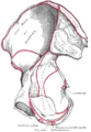
 Right hip bone. External surface.
 -
 Right hip bone. Internal surface.
 -
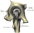 Left hip-joint, opened by removing the floor of the acetabulum from within the pelvis.
 -
 Hip bone.Medial view.
 -
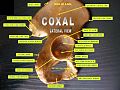 Hip bone. Lateral view.

.gif)


See also
[sửa | sửa mã nguồn]References
[sửa | sửa mã nguồn]Bài viết này kết hợp văn bản trong phạm vi công cộng từ trang , sách Gray's Anatomy tái bản lần thứ 20 (1918).
- ^ “The Hip Bone - Ilium - Ischium - Pubis - TeachMeAnatomy”. teachmeanatomy.info. Truy cập ngày 19 tháng 6 năm 2024.
- ^ a b c d Bojsen-Møller, Finn; Simonsen, Erik B.; Tranum-Jensen, Jørgen (2001). Bevægeapparatets anatomi [Anatomy of the Locomotive Apparatus] (bằng tiếng Đan Mạch) (ấn bản thứ 12). Munksgaard Danmark. tr. 237–239. ISBN 978-87-628-0307-7.
- ^ a b c Multiple citations to "(J Bridges)" embedded in text.Bản mẫu:Full citation
- ^ “Gray's Anatomy”. 1918. Bản gốc lưu trữ 22 Tháng mười hai năm 2009.
- ^ “hip fracture”. McGraw-Hill Concise Dictionary of Modern Medicine. 2002 – qua TheFreeDictionary.
- ^ "pelvimetry" tại Từ điển Y học Dorland
- ^ a b c d Romer, Alfred Sherwood; Parsons, Thomas S. (1977). The Vertebrate Body. Philadelphia, PA: Holt-Saunders International. tr. 188–192. ISBN 0-03-910284-X.
- ^ Dines, James P., et al. "Sexual selection targets cetacean pelvic bones." Evolution 68.11 (2014): 3296-3306.
External links
[sửa | sửa mã nguồn]| Wikimedia Commons có thêm hình ảnh và phương tiện truyền tải về Naazulene/Xương hông. |
- hip/hip%20bones/bones3 at the Dartmouth Medical School's Department of Anatomy